| THE RELATION OF THE INNER BORDER OF BACTERIAL FILM ON THE TOOTH WITHIN THE GINGIVAL CREVICE TO THE ZONE OF DISINTEGRATING EPITHELIAL ATTACHMENT CUTICLE* |
Charles C. Bass, M.D., New Orleans, La.
From the School of Medicine, Tulane University of Louisiana
Published September 1950.
*Studies promoted by facilities to which the author has had access at the School of Medicine. Tulane University of Louisiana, and by aid for equipment and supplies provided by the university.
|
IN THE original description of the zone of disintegrating epithelial attachment cuticle (zdeac),1 the statement was made that "in most places its course conforms to the course of the near-by border of the hard calculus found on such specimens."
The zdeac accurately indicates the location, before the tooth was extracted, of the outer border of the epithelial tissue (the epithelial attachment) attached to the tooth. Likewise, it indicates the extent to which the soft tissues have been removed from the surface of the tooth, in the course of the almost universal disease process, periodontoclasia.
By the inner border (or edge) of the calculus on a tooth is meant that part attached nearest to the zdeac and therefore nearest to the outer border of the living epithelial cells to the tooth. The purpose of this paper is to direct attention to the close relation of the inner border of subgingival calculus to the zdeac.
|
Material and Methods
Formalin-preserved specimens are selected. On teeth from young people
the calculus is usually located on the enamel and, therefore, above the cementoenamel junction line. On those from older persons, depending upon the progress that has been made by the disease process, often the calculus extends well below the cemento-enamel junction and then is located on the cementum. As the location of the zdeac moves apexward, usually calculus continues to build and follow close behind. The continuously changing location of the zdeac apexward is well illustrated in a recent paper2 which is recommended to those to whom this may not be entirely clear.
In general the same equipment and technical methods arc required as those originally described for demonstrating the zdeac. The specimen, without brushing or cleaning, is stained in crystal violet solution (O.5 per cent in water) for one-half to one minute. It is then brushed off (medium Masso brush) and rinsed in running water. It is now ready for examination while still wet, with the aid of incident lighting, under the dissecting microscope or under very low powers of the compound microscope. Such stained and brushed off specimens may be allowed to dry and are useful for further study, and for demonstration to others. Such dried specimens may be permanently mounted by placing the interesting side or area onto a coverglass and running a few drops of Clarite or balsam under it. Such mounted or unmounted stained and dried specimens can be kept for long periods of time. They are convenient and ready for demonstration to students or others.
|
 |
| Fig. 1 |
|
|
Observations
There is more or less variation in the width of the zdeac on different tooth specimens and at different locations around a given specimen. The apexward border of the disintegrating zone merges into the normal (not disintegrating) epithelial attachment cuticle. Therefore, there is no sharp line of demarcation between the affected and unaffected parts of this membrane.
The occlusalward border of the zdeac consists of cuticular material in varying stages of disintegration. There are loose particles, many of which are removed by the method of preparation suggested previously. In addition, there are partially detached particles, some of which also are broken off and removed by the brushing. Under any circumstance this side of the zdeac presents a ragged, irregular outline.
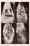
The tendency of the location of the zdeac and that of the inner border of subgingival calculus to conform to each other, and the close relation between them, may be illustrated by several photomicrographs taken of selected specimens. Fig. 1 shows the zdeac running somewhat diagonally across the root about one-third of the way down. Note that where the calculus projects farthest the zdeac gives way to it. At places the zdeac appears to project into gaps between advancing knobs of calculus.
The parallel relation of the zdeac and the inner border of the calculus scales on enamel is shown in Figs. 2 and 3. In Fig. 2 representing the condition in the fairly early stage of periodontoclasia, the calculus is thin and in patches. Note that the zdeac is about the same distance from, and follows the same general course as, the inner border of the calculus. At the far right side of the picture a scale of calculus projects into the zdeac. In Fig. 3 the calculus is thicker or heavier, and in places gives the impression that it was encroaching upon the zdeac. Note the gap in the calculus near the middle of the picture where the zdeac has not been forced downward quite so far.
|
 |
| Fig. 2, 3 & 4 |
|
|
In Fig. 4 the occlusalward part of the calculus lies over the cementoenamel junction. The zdeac dips, crescent-like, giving way to the advancing inner border of the calculus.
In Fig. 5 the calculus, most of which is below the cementoenamel junction, presents an irregular inner border, to the general outline of which the zdeac conforms, but it does not project into the indentations.
Fig. 6 shows the course of the zdeac following, in general, the course of the inner border of the calculus, closer in some places than in others. On the right side of the picture the heavy calculus had been cracked off in the manipulations of extraction.
In Fig. 7, where the periodontoclasia lesion extended as a pocket far down upon the side of the root, the course of the zdeac is seen to conform, in general, to the border of the advancing calculus. At some places knobs of calculus seem to project into the zdeac.
In Fig. 8 the zdeac runs from the cementoenamel junction at about midpoint diagonally halfway or farther along the root. Calculus which was present in the large, deep pocket on the labial side conforms, in general, to the course of the zdeac. Much of the deeper calculus consists of thin, irregular, patchy scales. On the other side the periodontal fibers are still present nearly to the cementoenamel junction, indicating that little or no destruction had occurred on this side. Note that the zdeac runs toward the incisal edge and is not satisfactorily visualized, in this specimen, above the cementoenamel junction.
|
 |
| Fig 5, 6, 7 & 8 |
|
|
Continue reading online CLiCK
|
|
|