| The Location of the Zone of Disintegrating Epithelial Attachment Cuticle |
| RELATION TO THE CEMENTO-ENAMEL JUNCTION AND TO THE OUTER BORDER OF THE PERIODONTAL FIBERS ON SOME TOOTH SPECIMENS |
by
Charles C. Bass, M.D.
and
Harold M. Fullmer, D.D.S.
Tulane University of Louisiana and
Charity Hospital of Louisiana
New Orleans, La.
St Louis Vol. 27, No. 5, Pages 623~628, October 1948
|
The epithelial attachment cuticle (eac) is a cuticular membrane which underlies the epithelial attachment (ea) and attaches the innermost cells of this tissue to the tooth, whether it is located on the enamel or on the cementum This cuticle is probably produced or laid down by the cells of the epithelial attachment.1–4
Wherever it is located at any time the outer border of the epithelial attachment cuticle is undergoing more or less breaking down or disintegration and as a result stains heavier than the remaining uninjured portion. This heavier staining line or strip may be demonstrated readily on extracted tooth specimens. Bass5 has called it the zone of disintegrating epithelial attachment cuticle (zdeac).
The outer border of the epithelial attachment comes right up to the zone of disintegrating epithelial attachment cuticle but does not overlap it. Therefore this line accurately indicates the location of the outer border of the epithelial attachment at any place around a given tooth specimen. It also accurately indicates the exact extent to which the epithelial attachment cuticle has been destroyed, and is now being destroyed, at any place.
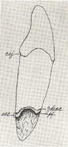
The purpose of this paper is to show the location of the zone of disintegrating epithelial attachment cuticle, the cemento-enamel junction (cej), and the outer border of the periodontal fibers (pi) on a number of selected tooth specimens.
|
MATERIAL AND METHODS
Specimens used for this purpose were selected from a large miscellaneous collection of formalin-preserved extracted teeth obtained from a number of different sources. An attempt was made to select representative specimens and not specimens showing unusual or rare conditions.
The method previously described5 was employed in preparation and examination of specimens. Any excessive adhering soft tissue was trimmed away. Undesirable (for this purpose) soft bacterial and cellular material was removed by brushing with a toothbrush. Vigorous brushing does not remove the zone of disintegrating epithelial attachment cuticle (it does remove some of the loose pieces at the outer border of the zone of disintegrating epithelial attachment cuticle), the epithelial attachment cuticle or the periodontal fibers. Most, if not all, adhering epithelial cells are removed by the brushing. The specimen was stained with crystal violet (0.5 per cent in H2O) one to three minutes and again brushed off gently.
Drawings were made with the aid of an Abbe type camera lucida (Zeiss) at approximately x112 original magnification. Outlines were accurately traced. The zone of disintegrating epithelial attachment cuticle blends or shades into the uninjured epithelial attachment cuticle; therefore the distinction between them is not usually as sharp as these pictures indicate. The outer side of the zone of disintegrating epithelial attachment cuticle, which consists of breaking up cuticular material5, is a more or less irregular line when viewed under considerable magnification. Notwithstanding these limitations, the drawings correctly indicate the general outline of the disintegrating zone.
The outline of the outer border of the periodontal fibers on the tooth was accurately drawn and the fibers at the border indicated. Fibers and periodontal tissue farther down on the root are suggested only by a few miscellaneous lines. An effort was made to trace and indicate accurately the outline of the root itself.
Calculus, bacterial film, and other material on the specimen above the zone of disintegrating epithelial attachment cuticle are not indicated (except in Fig. 1) neither are cavities nor other abnormalities.
Whenever the cemento-enamel junction was obscured by overlying calculus or other material, this material was removed to permit accurate tracing of the junction line (the cej) between the enamel and the cementum.
When a tooth is extracted, most of the tissue of the epithelial attachment is torn from it and remains in the mouth. The epithelial attachment cuticle, whether on the enamel or on the cementum, always remains firmly attached to the tooth. Usually a varying number of the epithelial attachment cells remain attached to the cuticle, and these may be recognized and identified by appropriate technique4. A convenient quick way is to stain (H & E or other stain) an extracted tooth specimen and examine it with suitable magnification under incident light. Higher magnification and transmitted light may be used to examine specimens of the epithelial attachment cuticle and the cells thereon, removed from the enamel with the aid of acid (10 per cent HCI in H2O). Paraffin or celloidin sections of decalcified teeth may be made for similar examination, including the epithelial attachment cuticle and any adhering cells on the cementum.
When a tooth is extracted, most of the periodontal fibers are torn, the dental end remaining firmly attached to the cementum. By suitable technique, the location of the outer border of these fibers on the tooth may be recognized satisfactorily. In this way one may observe exactly to what extent, from the cemento-enamel junction apexward, the fibers have been destroyed and have been replaced by epithelial attachment.
|
Changes that occur in the location of the epithelial attachment, epithelial attachment cuticle, zone of disintegrating epithelial attachment cuticle and periodontal fibers.
On a normal tooth, stabilized in normal occlusion, the epithelial attachment extends from the cemento-enamel junction occlusalward to the zone of disintegrating epithelial attachment cuticle. The underlying epithelial attachment cuticle extends from the cemento-enamel junction occlusalward to, and including, the zone of disintegrating epithelial attachment cuticle (Figs. 1, 2). Therefore the epithelial attachment, together with its underlying cuticle, is normally located on the anatomical crown.
Normally periodontal fibers cover the entire root from the cemento-enamel junction to the apex (Figs. 1, 2). An important function of the cementum is to imbed and anchor the dental ends of the different groups of these fibers to the tooth. As the outermost fibers break down or disintegrate and are removed from the surface of the cementum (in the usual course of the disease process, periodontoclasia), the epithelial attachment grows apexward, crossing over the cemento-enamel junction, and covering the area of cementum from which the periodontal fibers have been removed. At such a place the epithelial attachment is located partly on the enamel above, and partly on the cementum below the cemento-enamel junction (Figs. 3, 4). The occurrence of this condition frequently has been noted by others6,7,8 without, however, emphasizing its true pathological significance, except for Goldman6.
As the breaking down and removal of the periodontal fibers progresses further, the epithelial attachment grows farther apexward over the cementum from which the fibers have been removed. Usually, at this stage, the zone of disintegrating epithelial attachment cuticle and the outer border of the epithelial attachment recede at about the same rate as the inner border of the attachment advances along the tooth. In due course, the full width of the epithelial attachment and the zone of disintegrating epithelial attachment cuticle may be located entirely below the cemento-enamel junction at a given place around a tooth (Figs. 5-10). As the process continues, the epithelial attachment progressively moves apexward.
As the attachment grows apexward, simultaneously cuticle is produced beneath it. This attaches the new epithelial cells to the cementum. Therefore the epithelial attachment and its underlying cuticle extend from the zone of disintegrating epithelial attachment cuticle at any given place to the outer border of the remaining intact periodontal fibers.
|
 |
| Fig. 10 |
|
|
|
|
SUMMARY
The location of the zone of disintegrating epithelial attachment cuticle in relation to the cemento-enamel junction and to the outer border of the periodontal fibers on some extracted tooth specimens has been indicated by means of camera lucida drawings. The explanation of "continuous passive eruption" as a physiological process is questioned.
REFERENCES
1. Beeks, H.: Normal and Pathological Pocket Formation, J. A. D. A. 16: 2167, 1929.
2. Kronfeld, R.: The Epithelial Attachment and So-called Nasmyth's Membrane,
J. A.D. A. 17: 1907, 1930.
Orban, B.: Oral Histology and Embryology, St. Louis, 1944, The C. V. Mosby Company.
4. Gottlieb, B.: Der Epithelansantz Am Zahn, Deutsch. Monatschr. f. Zahnheilk. 39: 142, 1921.
5. Bass, C. C.: A Demonstrable Line on Extracted Teeth Indicating the Location of the Outer Border of the Epithelial Attachment, J. D. Res. 25: 401,1946.
6. Goldman, H.: The Relationship of the Epithelial Attachment to the Adjacent Fibers of the Periodontal Membrane, J. D. Res. 23: 177, 1944.
7. Orban, B., and Mueller, E.: The Gingival Crevice, J. A. D. A. 16: 1206,1929.
8. Skillen, W. G.: The Morphology of the Gingiva of the Rat Molar, J. A. D. .A. 17: 645,
1930.
Studies were promoted by facilities to which the authors have had access at the School of Medicine. Tulane University of Louisiana, and by aid for equipment and supplies provided by the University. Participation of the junior author was aided by the Veterans Administration assistance toward his work for the Masters degree.
Received for publication May 10, 1948.
|
|
|