The Bacterial Film
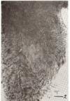
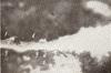
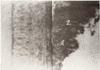
The bacterial film on the tooth is heavily stained by the one-minute exposure suggested previously. Wherever the film is very thick, the stain does not have time to soak into and stain the deeper part next to the tooth. However, the surface of the film pad, including the inner border of it at the zdeac, is well stained. At favorable places it can be seen that this film always extends right down to (Figs. 2 and 3) and often slightly overlaps (Figs. 4 and 5) the occlusalward border of the zdeac.
|
 |
| Fig. 4 |
|
|
 |
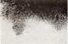
| Fig. 5 |
|
|
The characteristics and composition of this material can be observed best by picking a small particle from the inner border of the bacterial film and examining it under appropriate magnification. For most purposes it is only necessary to tease apart the particle in a small droplet of 50 per cent glycerine on a slide, put on a quarter-sized coverglass, and examine. Very fine-pointed teasing needles are required. Such manipulations arc best carried out under the dissecting microscope. Magnification of twelve to fifteen times is desirable for teasing out and preparing such material on the slide. Also, this material can be teased out in water on a slide, allowed to dry, and restained as desired.
Another good method for studying the nature of the inner border of the bacterial film and its relation to the zdeac is to select a favorable area where the zone is still on the enamel, dip in acid (10 per cent Hcl.) for a moment, remove a small piece of cuticle which includes the objects of interest, and restain this.
Sections made through appropriate areas on decalcified (10 per cent Hcl.) tooth specimens are also helpful in studying the bacterial film attached to cementum within the gingival crevice.
|
 |
| Fig. 6 |
|
|
 |
| Fig. 7 |
|
|
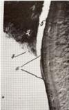
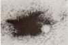
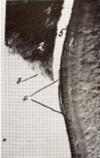
Material removed from the inner (apexward) border of the film pad approaching and overlapping the occlusalward border of the zdeac is found to consist mainly of parallel-arranged long rods and filaments. Growing and fruiting ends of these are usually found in preparations which include the surface and the extreme apexward feathery edge of the pad. This form was described by Beust3 in 1908 and designated by him as Leptothrix falciformis (Figs. 6 and 7). In 1929 he again directed attention to this organism in material from about the teeth, and pointed to the association of it with spirochetes.
From time to time, others5, 6, 7, 8, 9, 10, 11 have recognized what seems to be this same organism. Box9, 10 especially has shown convincingly, with photomicrographs of sections, that some of the fruiting heads tend to grow down-
*Reference 9, Figs. 1 and 2, reference 10, Fig. 4.
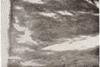
ward toward the very bottom of the pocket. Studies of large numbers of suitable preparations, as herein suggested, show that the film pad near the zdeac consists largely of parallel rods and filaments (Figs. 8 and 9), one end attached to the tooth and the other consisting mostly of growing ends and fruiting heads extending outward and downward into the open space of the gingival crevice. (Figs. 10, 11, 12, 13, and 14).
|
 |
| Fig. 8 |
|
|
 |
| Fig. 9 |
|
|
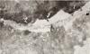
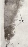
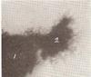
While this paper refers especially to attached bacterial film on the tooth at the bottom of the periodontal lesion, attention may be called to the great abundance and preponderance of spirochetes* in the narrow space between the attached film pad and the epithelial cell wall of the pocket. Spirochetes are so numerous at the very bottom of this space overlying the zdeac (Fig. 15) until, on favorable specimens and with good technique, one can pick material from over the zone, which consists of practically pure spirochetes (Fig. 16). The great abundance of spirochetes found upon and about the fruiting heads of leptothrix film is indicated in photomicrographs** published previously by me.12
*The term spirochete is used here in its broader sense to indicate spiral, corkscrew shaped organisms.
**Reference 12. Figs. 2 and 3.
|
 |
| Fig. 10 |
|
|
 |
| Fig. 11 |
|
|
Summary
The inner (apexward) border of the bacterial film on the tooth within the gingival crevice extends right to and usually overlaps the outer (occlusalward) border of the zdeac. The attached bacterial film consists of a pack of more or less parallel long rods and filaments, one end attached to the tooth and the other extending outward (from the tooth) into the narrow space between this film and the crevicular epithelium.
There is a great abundance of spirochetal organisms closely associated with the growing ends and fruiting heads in this space, especially the space immediately over the zdeac.
|
 |
| Fig. 15 |
|
|
 |
| Fig. 16 |
|
|
References
1. Bass, C. C.: A Demonstrable Line on Extracted Teeth Indicating the Location of the Outer Border of the Epithelial Attachment, J. D. Res. 25: 401,1946.
2. Bass, C. C., and Fullmer, H. M.: The Location of the Zone of Disintegrating Epithelial Attachment Cuticle in Relation to the Cemento-Enamel Junction and to the Outer Border of the Periodontal Fibers on Some Tooth Specimens, J. D. Res. 27: 623, 1948.
3. Beust, Theo,: A Contribution to the Morphology of the Microorganisms of the Mouth, Dental Cosmos 50: 594, 1908.
4. Beust, Theo.: Spirochetes and Fusiform Bacilli of the Mouth, J. Am. Dent. A. 16: 1415,1929. (Sec also J. Am. Dent. A. 16: 714.)
5. Goodrich, H. P., and Mosely, M. ,J.: Certain Parasites of the Mouth in Cases of Pyorrhoea, J. Roy. Micr. Soc. 61: 513,1916.
6. Aisenberg, M. S.: Morphologic Studies of Microorganisms of Fusiform Type, Dental Cosmos 75: 546, 1933.
7. Dean, R. D., and Dean, M. T.: Cultural Observations on Fusospirochetal Infections, J. D. Res. 11: 759, 1931.
8. Tunnicliff, R.: Fusiform Organisms in the Mouth, J. D. Res. 17: 53, 1938.
9. Box, n. K.: Can Specific Infective Factors Operate to Deepen a Pocket? J-.Canadian D. A. 10: 427, 1944.
10. Box, H. K.: New Aspects of Periodontal Research, J. Canadian D. A. 13: 3, 1947. 11. Cobe, H. M.: Vincents Infection: Experimental Reproduction of Lesions and the Role of Streptococci, J. Am. Dent. A. 37: 317,1948.
12. Bass, C. C.: Habitat of Endameba Buccalis in Lesions of Periodontoclasia, Proc. Soc. Exper. Biol. & Med. 66: 9,1947.
|
Return to prev. page CLiCK
|
|
|